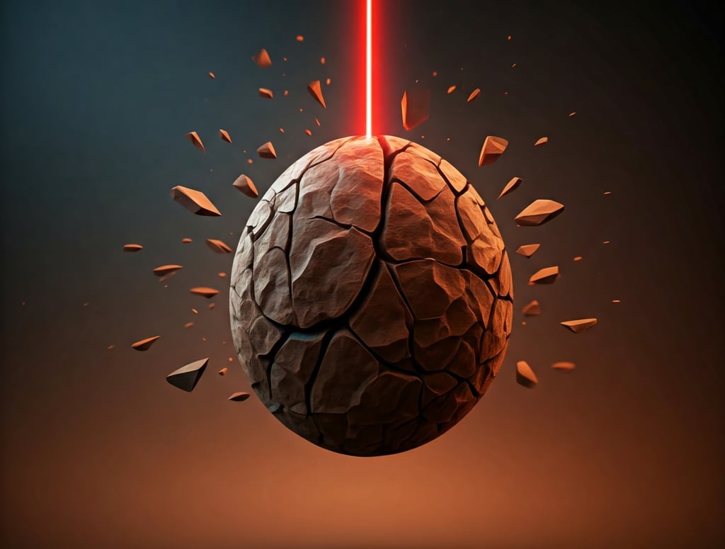
What size of kidney stone requires surgery? (mm Size Guide)
Stones over 6–7mm often won't pass; almost all over 10mm usually need treatment. A consultant urologist explains how size, location and type decide if you need surgery.
URINARY STONE DISEASE
Ivo Dukic, Private Urologist and kidney stone surgeon
12/8/20243 min read
Written and Medically Reviewed by Mr Ivo Dukic, Consultant Urologist | Last updated: 16/03/2026
Introduction to Kidney Stones
Kidney stones, also known as renal calculi are usually hard dense crystals that form inside the kidneys. Their formation is a complex process that often results from an imbalance in the substances that are filtered through the kidneys. Factors such as dehydration, dietary choices, and underlying health conditions can contribute significantly to the formation of these stones. There are various types of kidney stones which are made from a variety of minerals. The most common type of kidney stones are calcium oxalate stones.
Stone types
Non-infection stones (1)
• Calcium oxalate
• Calcium phosphate
• Uric acid
• Ammonium urate
Infection stones (1)
• Magnesium ammonium phosphate
• Highly carbonated apatite
• Ammonium urate
Genetic causes (1)
• Cystine
• Xanthine
• 2,8-Dihydroxyadenine
While spontaneous passage of kidney stones and ureteric stones is possible for most smaller kidney stones, surgical intervention may be necessary. This article outlines the factors considered in determining the need for surgery, with a focus on the guidelines of the European Association of Urology (EAU) and the National Institute for Health and Care Excellence (NICE).
What size of kidney stone requires surgery?
Stone size is a critical factor, with larger stones having a lower likelihood of spontaneous passage and an increased risk of complications such as obstruction and infection.
EAU and NICE Guidance: Both organizations generally recommend surgical consideration for stones exceeding 10 millimeters (mm) in diameter as these are unlikely to pass. However, this is not an absolute threshold. Stones that are greater than 5 to 7 mm will often not pass by themselves and if blocking the ureter will cause significant pain and or blockage of the kidney system.
Other factors influencing the decision to pursue surgery include:
Stone Location: Stones located in the upper ureter (the tube connecting the kidney to the bladder) may have a higher probability of spontaneous passage compared to those situated in the lower ureter or near the bladder.
Kidney anatomy - kidneys vary in shape, size, rotation and position. Not all treatments are suitable for all patients as some kidneys will not drain normally.
Stone Composition - depending on your predisposition to forming further stones and any underlying health factors. Certain stones such as uric acid stones and infection stones respond less well to shockwave treatment.
Patient-Related Factors: Individual patient characteristics, such as age, overall health status, and pain tolerance, are considered. Patients with underlying medical conditions or a low pain threshold may be more likely to benefit from surgical intervention.
History of Previous Episodes: Individuals with a history of kidney stones or recurrent episodes may be considered for more proactive treatment, even for smaller stones.
Surgical Options for Kidney Stone Management
Several surgical procedures are available for the removal of kidney stones:
Shockwave Lithotripsy (ESWL): This non-invasive technique utilizes high-energy sound waves to fragment the stone into smaller pieces that can be passed naturally. It is generally suitable for stones smaller than 10 mm to 20 mm but may require multiple treatments.
Ureteroscopy: This minimally invasive procedure involves inserting a thin, flexible tube (ureteroscope) through the urethra and bladder into the ureter to access and remove the stone. It is effective for a range of stone sizes and locations but is usually recommended for kidney stones smaller than 15 to 20 mm.
Percutaneous Nephrolithotomy (PCNL): This procedure involves creating a small incision in the back to directly access the kidney and remove the stone. It is typically used for larger stones, greater than 15 mm or those that are difficult to reach with other methods.
When to Seek Medical Attention
Individuals experiencing symptoms such as severe flank pain, blood in the urine, nausea, or vomiting should consult a healthcare professional promptly. Early diagnosis and appropriate treatment can help prevent complications and facilitate a faster recovery.
Conclusion
The decision regarding surgical intervention for kidney stones requires a comprehensive assessment of various factors. While stone size is a significant consideration, the EAU and NICE guidelines emphasize a multi-faceted approach that accounts for individual patient characteristics, stone location, composition, and previous medical history. Consultation with an endourologist (a urologist specialising in kidney stone surgery) is crucial to determine the most appropriate treatment plan for each case.
Mr Ivo Dukic is an experienced consultant urologist and high-volume stone surgeon in Birmingham, United Kingdom. He is a leading expert in complex kidney stone surgery who offers personalised consultations in Birmingham, United Kingdom. He regularly performs extracorporeal shockwave lithotripsy (ESWL), ureteroscopy and laser lithotripsy, and percutaneous nephrolithotomy (PCNL) using the very latest technology.
Schedule an appointment with him for expert, bespoke advice through his Top Doctors profile or book an appointment through Harborne Hospital, HCA Healthcare, the Priory Hospital, Edgbaston, Circle Health Group or Droitwich Spa, Circle Health.
References
European Association of Urology. Uroweb. Urolithiasis. Available at: https://uroweb.org/guidelines/urolithiasis. Accessed November 29, 2024.
National Institute for Health and Care Excellence. Kidney stones and bladder stones: diagnosis and management. Available at:
https://www.nice.org.uk/guidance/ng118. Accessed November 29, 2024.
Ivo Dukic
Contacts
e-mail: admin@ivodukic.co.uk
Telephone number for private patients:
0121 716 9046
(Mondays to Fridays 0800 - 18:00)
For NHS patients seen at University Hospitals Birmingham NHS Hospitals please get in contact on
0121 424 9011
(Mondays to Fridays 0900 - 17:00)
ivodukic.co.uk
© 2026 UrolSurg LTD


GMC 6103172
BSc MBChB PG Cert FRCSEd (Urol)