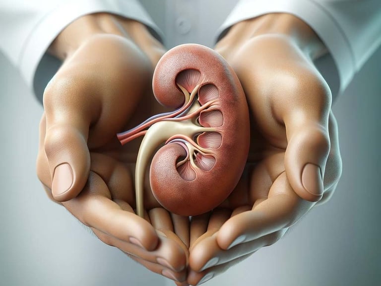
Ureteroscopy for kidney stone disease
CONDITIONS


Introduction
Ureteroscopy is a common procedure used to treat kidney stone disease. Kidney stones form inside the kidney and can cause problems whilst in the kidney or if they drop into and block the tube draining the kidney, the ureter. The ureter is the tube connecting the kidney to the bladder. A ureteroscopy operation is a minimally invasive procedure and is an often chosen method for managing ureteric stones and kidney stones due to its effectiveness and relatively low-risk profile. This article will explore the indications, benefits, risks, and follow-up care associated with a ureteroscopy procedure.
Indications for Ureteroscopy
Ureteroscopy is indicated for patients with ureteric stones or kidney stones that are unlikely to pass spontaneously or are causing significant symptoms. Specific indications include:
Persistent Pain: When patients experience ongoing pain despite conservative management of kidney stones (letting the stones pass naturally) ureteroscopy may be recommended.
Obstruction: Stones obstructing the urinary tract may lead to potential kidney damage; this may necessitate intervention
Infection: In cases where stones are associated with urinary tract infections, prompt removal is essential to prevent complications occasionally this first requires a ureteric stent insertion or a tube in the kidney (nephrostomy)
Failed medical therapy: When medical expulsive therapy fails to facilitate stone passage, ureteroscopy becomes a viable option
Stone size and location: Stones larger than 5-7 mm or located in the upper ureter are less likely to pass on their own and often require ureteroscopy. Stones larger than 10-15 mm may require either a ureteroscopy procedure or a PCNL operation.
Benefits of Ureteroscopy
Ureteroscopy offers several benefits over other treatment modalities:
High Success Rate: Ureteroscopy has a high success rate in stone removal, with most stones being completely removed in a single session (70-90% depending on location, number and size of stones).
Minimally Invasive: The procedure is performed using a small, flexible scope, minimizing tissue damage and reducing recovery time
Immediate Relief: Patients often experience immediate relief from symptoms such as pain and obstruction following the procedure
Versatility: Ureteroscopy can be used to treat stones of various sizes and locations within the ureter
Diagnostic Capability: The procedure allows direct visualization of the urinary tract, aiding in the diagnosis of other potential issues
Risks of Ureteroscopy
While ureteroscopy is generally safe, it is not without risks. Potential complications include:
Infection: There is a risk of urinary tract infection following the procedure, which may require antibiotic treatment
Bleeding: Minor bleeding is common, but significant bleeding is rare
Ureteral Injury: The ureteroscope can cause injury to the ureter, leading to strictures or perforation. Very rarely this requires and open operation to fix the injury.
Stone Migration: Stones may move to a different location during the procedure, complicating removal
Failure to clear the stones: Some stones cannot be cleared with a ureteroscope due to the internal angles inside the kidney or due narrowing of the ureter or impaction of stones in the ureter.
Ureteric stent symptoms: Ureteric stent often affect your ability to work and are associated with urinary symptoms, abomdinal pain, blood in the urine, rushing to get to the toilet and urinary tract infections.
Anesthesia Risks: As with any procedure requiring anesthesia, there are associated risks, particularly in patients with underlying health conditions
You can find more information about the indications and risks from the British Association of Urological Surgeons. There is also further information on kidney stones provided by the European Association of Urology.
Animated video from the European Association of Urology (below)

Following the procedure:
Remember, successful stone management extends beyond the procedure itself:
Hydration: Aim for 2.5 -3 litres of water daily to prevent or delay further stone formation.
Dietary modifications: Minimize stone-forming foods like animal protein and oxalates based on your stone type.
A ureteric stent is often inserted with some removal strings which come outside the urethra. The ureteric stent is often removed 3-5 days following a ureteroscopy operation. In some cases a ureteric stent can be left in for a few weeks to allow healing of a ureter if there are problems during your procedure. For these patients a further procedure where a flexible camera is inserted under local anaesthetic into the bladder is required to remove the ureteric stent.
Scheduled check-ups can ensure timely detection and management of any recurrent stones if required.
After surgery if you are having ongoing pain, urinary tract infections of blood in the urine please get in contact with your surgeon so that we can organise further imaging tests (usually a CT scan) to look for any problems following the surgery.
This article provides general information and should not substitute for professional medical advice that is tailored to your situation with your urologist. Always consult a urologist for a personalised assessment and treatment plan.
The specific risks and benefits of ureteroscopy may vary depending on individual circumstances and your exact presentation.
Mr Ivo Dukic is regarded as one of the best kidney stone surgeons in the UK, combining vast surgical experience in kidney stone surgery with a patient-centred approach from his base in Birmingham. His practice is a destination for patients seeking definitive treatment for all types of kidney stone disease.
Why Patients Choose Mr Dukic for their kidney stone surgery:
Expert in Complex Kidney Stone Surgery: Possesses the specialist skill required to manage large, complex, and recurrent kidney stones that other surgeons may not be equipped to treat.
High-Volume PCNL Surgeon: As a high-volume surgeon, he performs a significant number of PCNL procedures annually, a key indicator of proficiency and one of the reasons he is considered amongst the best kidney stone surgeons in the UK.
Pioneer in Minimally Invasive Treatment: Utilises the most advanced, state-of-the-art techniques, including supine PCNL, mini-PCNL and ultra-mini PCNL, to minimise patient recovery time and improve surgical outcomes.
Accessible Across the UK: While based in Birmingham, he treats patients who travel from the West Midlands and across the entire United Kingdom for his specialist care.
You can schedule an appointment with him for expert, bespoke advice through his Top Doctors profile or book an appointment through Harborne Hospital, HCA Healthcare, the Priory Hospital, Edgbaston, Circle Health Group or Droitwich Spa, Circle Health.
Have any questions?


If you have any questions or wish to make an appointment please get in contact.
Ivo Dukic
Contacts
e-mail: admin@ivodukic.co.uk
Telephone number for private patients:
0121 716 9046
(Mondays to Fridays 0800 - 18:00)
For NHS patients seen at University Hospitals Birmingham NHS Hospitals please get in contact on
0121 424 9011
(Mondays to Fridays 0900 - 17:00)
ivodukic.co.uk
© 2026 UrolSurg LTD
GMC 6103172
BSc MBChB PG Cert FRCSEd (Urol)


